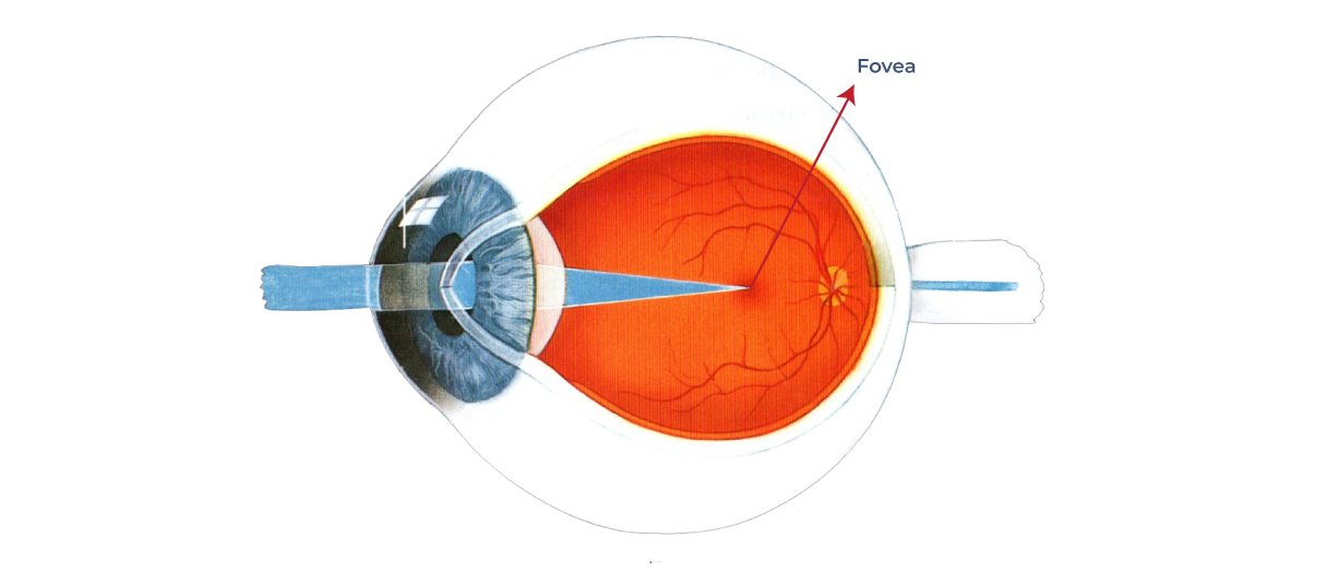
The fovea is a small but critical part of the human eye that plays an essential role in our ability to see fine details and perceive color. It is a tiny depression in the retina, located at the center of the macula, which is the central part of the retina responsible for our central vision. We often explain that if the retina were the size of the United States, the macula may be the size of one or two Midwest states, and the fovea would be the size of a few counties.
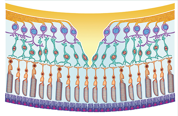
The fovea is only about 1.5 mm in diameter, but it contains an incredibly high concentration of photoreceptor cells called cones, which are responsible for detecting color and fine details in our visual environment. This high concentration of cones in the fovea makes it the most sensitive part of the retina and is responsible for our ability to see sharp images of objects that we are looking directly at.
The fovea is also unique in the way it is organized. The cones in the fovea are packed so tightly together that they are almost touching, which allows for an incredible level of detail and clarity in our central vision. In addition, the fovea is surrounded by a special layer of cells called the macular pigment, including Lutein and Zeaxanthin, which protects it from harmful UV rays and helps to improve contrast sensitivity and reduce glare.

The fovea is a small but critical part of the human eye that plays an essential role in our ability to see fine details and perceive color. It is a tiny depression in the retina, located at the center of the macula, which is the central part of the retina responsible for our central vision. We often explain that if the retina were the size of the United States, the macula may be the size of one or two Midwest states, and the fovea would be the size of a few counties.
Despite its small size, the fovea is an essential part of our visual system and plays a critical role in our daily lives. Any damage or degeneration of the fovea can significantly impact our central vision, making it difficult to read, drive, or perform other essential tasks that require fine detail vision.
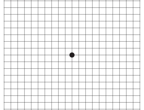
We often explain to patients that the retina is like a dart board and the fovea is like the bullseye of this dart board. Differing values of points correspond to each area of the dart board in the same way that different areas of the retina correspond to areas of our vision. With its high concentration of light-sensing cells and very small area compared to the much larger retina, the fovea is like the bullseye of the dart board, providing the highest value of “points” and fine detail of our center vision. This is why patients with damage to the fovea have corresponding blurriness, distortion, and even missing areas in their central vision, but essentially normal peripheral vision. Conditions such as age-related macular degeneration, central serous chorioretinopathy, and cystoid macular edema are just a few of these conditions that primarily affect the fovea.
In summary, the fovea is a small, highly specialized area of the retina that is responsible for our central vision and allows us to see fine details and perceive color. It is an incredibly important part of our visual system and requires careful attention and care to maintain optimal visual function.


Leave A Comment