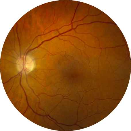
What is an epiretinal membrane?
Epiretinal membranes are fine sheets of fibrous tissue on the surface of the retina, which, similar to a scar elsewhere on your body, may contract and change the shape of the surrounding tissue. When an epiretinal membrane develops on the surface of the retina, the resulting changes to the delicate retinal tissue, can cause decreased vision and distorted or wavy lines in the central vision. Epiretinal membranes are also known as “macular pucker,” due to the puckered appearance of the retinal tissue and the resultant swollen, “puckered” appearance of the macula, with loss of the normal foveal depression.
What is the cause of epiretinal membranes?
The occurrence of epiretinal membranes is associated with prior inflammation, retinal detachment, retinal laser or cryotherapy, or trauma. Most often, epiretinal membranes occur spontaneously and without a clearly identifiable cause.
What is the treatment for epiretinal membranes?
Most patients with epiretinal membranes have mild symptoms and periodic examinations with an optometrist or ophthalmologist is recommended. If a decrease in central vision or symptomatic distortion develops in the setting or an epiretinal membrane, and no other clear cause can be identified, a surgical evaluation may help determine if surgery may be indicated. Vitrectomy, the surgical removal of the vitreous gel from the back of the eye, followed by peeling of the epiretinal membrane from the macula surface, is the preferred treatment for epiretinal membranes/macular pucker. While the visual acuity following surgery for epiretinal membrane rarely returns to perfectly normal, 20/20 vision, surgery improves the vision in approximately 75% of cases, typically decreases the visual distortion, and halts further progression of the epiretinal membrane.

