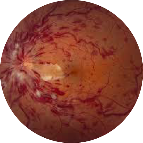
Retinal vein and arterial occlusions
The eye, like other organs of the body, has an arterial blood supply that delivers oxygen and other nutrients to the retina, and a venous blood drainage system which removes carbon dioxide and other waste products from the tissue, returning the blood to the heart and then to the lungs for oxygen replenishment and systemic circulation. A blockage in the retinal arteries, is similar to a stroke, and often results in irreversible damage to retinal tissue and permanent vision loss. If the central retinal artery is occluded, this is known as a central retinal artery occlusion, and if a branch off of the central retinal artery is affected, this is known as a branched retinal artery occlusion. A blockage of the retinal venous drainage results in leakage of fluid and blood, resulting in swelling of the affected retina. If the central retinal vein is occluded, this is known as a central retinal vein occlusion, and if a branch off of the central retinal vein is occluded, this is known as a branched retinal vein occlusion.
What is the treatment for a central retinal artery occlusion or branched retinal artery occlusion?
Unfortunately, there is no clinically proven treatment for central retinal artery occlusions, though several therapies have been attempted, including hyperventilation, paracentesis, ocular massage, or lowering of intraocular pressure. Individuals with an acute-onset central or branched retinal artery occlusion should be referred urgently to a nearby stroke center for a stroke work-up, which should include blood tests, EKG, brain imaging (MRI preferred, but head CT also acceptable), and a CTA, MRA, or ultrasound of the carotid arteries. If no cause can be identified, an echocardiogram should also be obtained.
What is the treatment for a central retinal vein occlusion or branched retinal vein occlusion?
Inasmuch as vision loss related to retinal vein occlusions is primarily due to swelling of the macula, your retina specialist may inject an anti-VEGF medication (Avastin, Eylea, or Lucentis) or a steroid medication (Ozurdex, Iluvien) into the back of the eye. If abnormal blood vessel growth occurs, laser photocoagulation may be used to decrease the risk of vitreous hemorrhage and retinal detachment. Your primary care physician should be involved to help screen and manage known risk factors of retinal vein occlusions, including systemic hypertension, smoking, diabetes mellitus, high cholesterol, and other systemic conditions.

