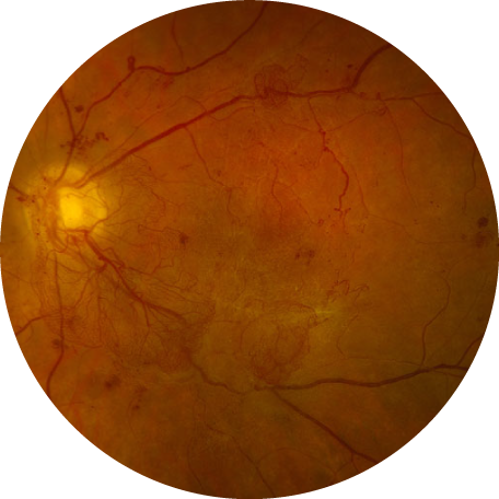
What is Diabetic Retinopathy?
Diabetic retinopathy occurs when high blood sugar levels cause damage to blood vessels in the retina. These blood vessels can swell and leak, they can close – stopping blood from passing through, or can lead to abnormal new blood vessels growth from within the retina into the vitreous gel. These various blood vessel changes, are collectively known as diabetic retinopathy, and unless appropriately treated, can lead to permanent vision loss and even blindness.
Do all patients with diabetes develop diabetic retinopathy?
Many large studies have examined the progression and treatment of diabetic retinopathy. The Wisconsin Epidemiological Studies found that the development of diabetic retinopathy depends largely on how long a patient has had diabetes mellitus, with longer duration of disease being associated with increased risk of diabetic retinopathy. In fact, nine out of ten patients who have had diabetes mellitus for 17-25 years develop diabetic retinopathy. However, not just duration of diabetes, but also recent blood sugar control, are associated with diabetic retinopathy. Each visit to our office, we will ask you what your most recent Hgb a1c level is, as this measure helps estimate blood sugar levels for the past several months and helps us estimate your risk for developing various eye problems seen in diabetic retinopathy.
How does diabetic retinopathy cause vision loss and blindness?
Diabetic retinopathy causes vision loss through several mechanisms, including the following:
leakage of proteins and cholesterol through small, damaged blood vessels. This leakage causes swelling (“edema”) in the macula, the area of your retina which corresponds to your central vision. This is known as diabetic macular edema.
The damage to the small blood vessels in the macula can lead to complete closure of these blood vessels, and thus preventing blood flow (“perfusion”) to the corresponding retina. If this blockage and “non-perfusion” occurs in your macula, your vision could be affected.
In an effort to improve compromised blood flow to the retina, the body creates new blood vessels, which start within the retina (the inner lining of the back of the eye). These abnormal blood vessels can grow, like vines of weeds in your garden, from the retina into the vitreous gel which fills the back of the eye. When this vitreous gel moves, the abnormal blood vessels can bleed into the vitreous gel. This is known as a diabetic vitreous hemorrhage.
These abnormal blood vessels (“neovascularization”) growing into the vitreous gel remain attached to the retina, which can cause a tractional, tugging force on the retina, which can cause the retina to separate from the eye wall. This is known as a diabetic tractional retinal detachment.
My doctor says I have severe, non-proliferative diabetic retinopathy and I am at risk for developing proliferative diabetic retinopathy. What does this mean? What are the various stages of diabetic retinopathy?
Diabetic retinopathy is graded using various grading and classification criteria. These criteria help estimate risk of developing vision loss related to diabetes, and help dictate how frequently you need a dilated eye exam to appropriately screen for signs of retinopathy and receive appropriate treatment to restore or prevent vision loss. Click here for a summary of the grading and classification fo diabetic retinopathy. In general, patients with mild, non-proliferative diabetic retinopathy should be followed at least every 12 months, those with moderate or severe non-proliferative diabetic retinopathy should be followed every 6-12 months, and those with proliferative diabetic retinopathy should be seen every 3-4 months.

What is the treatment for diabetic macular edema and proliferative diabetic retinopathy?
Swelling in the retina due to diabetes is treated either with injections of medicine into the eye, or with laser. Injections of medicine into the vitreous gel in the back of the eye, known as intravitreal injections, are the most common procedure performed by retina specialists. The following medications are all used for treatment of diabetic macular edema: Bevacizumab (Avastin), Ranibizumab (Lucentis), Aflibercept (Eylea), dexamethasone (Ozurdex) and fluocinolone acetonide (Iluvien). Several of these medications can also be used for the abnormal blood vessel growth seen in diabetes. Laser to the macula (focal grid laser) is also often used for diabetic macular edema, and laser to the peripheral retina (panretinal photocoagulation) is a mainstay for prevention of long-term complications of proliferative diabetic retinopathy.
At what point does diabetic retinopathy need treatment?
Diabetic retinopathy typically needs treatment when vision is affected, or at risk of being affected by swelling in the retina (diabetic macular edema) or when new blood vessel growth occurs (proliferative diabetic retinopathy) that has led to, or could lead to, diabetic vitreous hemorrhage or retinal detachment.

